
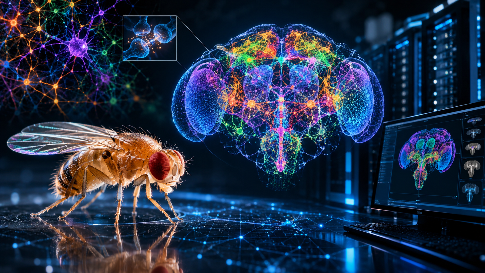
When Neo awakens wired into bioelectric cables in The Matrix (1999), or when entire human consciousnesses are transferred into cloud servers in Black Mirror’s San Junipero and the virtual worlds of Sword Art Online, science fiction flirts with the Holy Grail of neuroengineering: Whole Brain Emulation (Mind Uploading). On screen, the theoretical premise has always seemed straightforward — if the mind is the physical product of electrical spikes and chemical neurotransmitters traversing a finite network of neurons, scanning every synaptic connection and running that circuit inside a digital physics engine should recreate the behavior of the biological organism in silicon.
For decades, this vision collided with an insurmountable technological chasm. Until recently, humanity had only managed to fully map the nervous systems of microscopic, rudimentary creatures such as the nematode worm Caenorhabditis elegans (with a mere 302 neurons mapped in 1986). While the technology industry celebrated Large Language Models (LLMs) built on abstract algebraic matrices that consume megawatts of electrical power, a humble fruit fly (Drosophila melanogaster) continued executing acrobatic flight maneuvers, spatial navigation, decision-making, and olfactory memory using a poppy-seed-sized brain running on fractions of a microwatt.
That historical barrier has officially collapsed. In a landmark scientific milestone published across a special package of nine papers in Nature, the international FlyWire Consortium — led by Mala Murthy and Sebastian Seung at Princeton University alongside collaborators at the University of Cambridge — completed the first neuronal wiring diagram of an adult brain, mapping exactly 139,255 neurons and 54.5 million synapses. Even more remarkably, a team led by Philip Shiu demonstrated in Nature a computational whole-brain model of Drosophila, which has now been coupled to the virtual biomechanical skeleton NeuroMechFly v2 and integrated in closed-loop simulations inside the MuJoCo physics engine (arXiv:2602.17997) as well as commercial demonstrations by Eon Systems. For the first time in human history, the real connectome of a complex animal has come alive inside a computer.
1. The Chasm Between Artificial Neural Networks and the Biological Connectome
Why a Fruit Fly Brain Humbles AI Supercomputers
In contemporary discussions around Artificial Intelligence, it is common to hear that modern models are “brain-inspired.” In computer engineering practice, that comparison is skin-deep. A modern Transformer (such as GPT-4 or Claude) operates through homogeneous layers of floating-point matrix multiplications, where each “artificial neuron” is merely a static mathematical function lacking internal temporal dynamics, requiring liquid-cooled GPU farms just to predict the next token in a sentence.
The biological brain of Drosophila melanogaster, by contrast, is a masterpiece of evolutionary neuromorphic computing. Packed into a volume of just 0.03 cubic millimeters (roughly 750 × 350 × 250 micrometers), evolution compressed 139,255 highly specialized neurons divided into more than 8,453 distinct cell types. Unlike an LLM — where network topology is generic and weights are trained from scratch via statistical brute force — the fly connectome is born with a geometric architecture refined over hundreds of millions of years of natural selection.
From the C. elegans Worm to the Exponential Leap of Drosophila
To grasp the engineering magnitude of the FlyWire project, one must examine the logarithmic scaling of structural connectomics over the past four decades. Mapping a brain at synaptic resolution requires resolving structures at the nanometer scale, where visible light fails due to the optical diffraction limit.
| Organism / Project | Milestone Year | Total Neurons | Mapped Chemical Synapses | Raw Imaging Data (EM) | Behavioral Complexity |
|---|---|---|---|---|---|
| C. elegans (Nematode Worm) | 1986 | 302 | ≈ 7,000 | < 1 Gigabyte (Physical micrographs) | Simple tactile reflexes and undulatory locomotion |
| Drosophila Larva (1st Instar) | 2023 | 3,016 | ≈ 548,000 | ≈ 1 Terabyte | Crawling, chemotaxis, and basic associative learning |
| Adult Drosophila (Janelia Hemibrain) | 2020 | ≈ 25,000 | ≈ 20 million | ≈ 26 Terabytes | Partial central brain processing |
| Complete Adult Drosophila (FlyWire) | 2024–2026 | 139,255 | 54.5 million | ≈ 106 Terabytes | 3D flight, compound vision, navigation, courtship, and decision-making |
| Adult Mouse (Future Target) | Projected (2030+) | ≈ 70 million | ≈ 70 billion | ≈ 1 Exabyte (1,000 TB) | Full mammalian cognition and episodic memory |
The leap from the 302 neurons of C. elegans to the 139,255 neurons of the adult Drosophila is not merely a 460-fold numerical increase; it represents a phase transition in graph complexity. While the worm crawls in two dimensions, the fly commands an articulated exoskeleton with six multi-jointed legs, wings beating at 200 Hertz, compound eyes, and hypersensitive olfactory antennae.
2. The Engineering of FlyWire: Slicing a Brain into 7,062 Nanometric Layers
Transmission Electron Microscopy (TEM) at 4 Nanometers
No magnetic resonance scanner or clinical CT machine can resolve a synapse. The axons and dendrites that form the wiring of the fly brain narrow down to diameters of just 40 to 50 nanometers. To capture this wiring without losing a single cable, the project built upon the FAFB (Full Adult Fly Brain) dataset acquired at the Howard Hughes Medical Institute’s Janelia Research Campus.
The physical acquisition pipeline operates at the absolute limit of materials science:
- Chemical Fixation and Heavy-Metal Staining: The brain of an adult female Drosophila is preserved and stained with osmium, uranium, and lead atoms, which bind to lipid membranes and synaptic vesicles to scatter incoming electrons.
- Serial Ultramicrotomy with a Diamond Knife: The solid resin block containing the brain was mechanically sliced by an ultra-sharp diamond blade into exactly 7,062 serial sections, each only 40 nanometers thick (roughly 2,000 times thinner than a human hair).
- High-Throughput Electron Beam Scanning: Every slice was imaged via Transmission Electron Microscopy at a lateral resolution of 4 × 4 nanometers per pixel, generating a three-dimensional cube of over 100 Terabytes of grayscale imagery.
3D Convolutional Neural Networks and Global Human Proofreading
Having 7,062 stacked photographs does not automatically yield a brain map. In raw electron microscopy images, neurons appear as tangled gray blobs resembling a microscopic plate of spaghetti. If a single human researcher attempted to manually trace the contours of all 139,255 neurons across thousands of slices, the task would take more than 4,000 person-years.
Sebastian Seung’s laboratory at Princeton broke this bottleneck by deploying 3D Convolutional Neural Networks (3D CNNs) specialized in membrane boundary detection and Flood-Filling Networks. The AI swept across all 106 Terabytes to automatically segment the 3D volume of each neuron, while convolutional detectors pinpointed 54.5 million synaptic clefts and machine-learning classifiers predicted which of the six primary neurotransmitters each neuron releases: acetylcholine (the primary excitatory transmitter in insects), GABA (inhibitory), glutamate (excitatory or inhibitory in flies), dopamine, serotonin, or octopamine.
Even state-of-the-art AI, however, commits occasional false merges (mergers) or false splits (splits) when a 40 nm slice suffers a microscopic fold or dust artifact. That is where FlyWire’s collaborative cloud architecture stepped in, uniting hundreds of neuroscientists across more than 140 laboratories worldwide alongside trained citizen scientists (veterans of the Eyewire project). Together, humans and algorithms executed over 3 million manual proofreading edits to deliver a 100% audited connectome comprising 149.2 meters of biological wiring packed inside a head less than one millimeter wide.
3. From Static Map to Dynamic Mind: Philip Shiu’s LIF Model
The Mathematics of the Leaky Integrate-and-Fire (LIF) Neuron
A road map is not traffic. While Dorkenwald et al. delivered the static topology (who connects to whom and with how many synapses), proving that this wiring diagram contained the functional logic of the animal required Philip Shiu, Gabriella Sterne, and Mala Murthy to transform the static graph into an executable dynamic brain.
In their companion study published in Nature (DOI: 10.1038/s41586-024-07763-9), the researchers instantiated all 139,255 neurons inside the Brian2 neural simulator as biophysical Leaky Integrate-and-Fire (LIF) computational units. The electrical physics of every digital neuron follows real electrophysiological parameters:
- Resting Potential (V_rest): Held at -52 mV, reflecting the natural ionic equilibrium across the fly’s biological cell membrane.
- Firing Threshold (V_th): Fixed at -45 mV. When incoming synaptic currents push the membrane voltage across this threshold, the neuron fires an action potential (spike).
- Refractory Period and Time Constant: After firing, voltage resets and the neuron remains unresponsive for 2.2 milliseconds, governed by a membrane time constant (τ_m) of 5 milliseconds and a synaptic propagation delay of 1.8 milliseconds.
- Automated Chemical Polarity: Every connection weight was scaled strictly by the exact number of synaptic contacts counted under the electron microscope, multiplied by the sign of the predicted neurotransmitter (+1 for excitatory cholinergic synapses, -1 for inhibitory GABAergic and central glutamatergic synapses).
Predicting Real Behavior Without a Single Line of “If / Else” Code
The outcome stunned the neuroscience community. Without programming a single explicit behavioral rule and without training the network via backpropagation, the researchers simply stimulated the virtual sensory neurons corresponding to sugar touching the fly’s proboscis (gustatory receptor neurons, GRNs).
The cascade of electrical spikes propagated through the hidden layers of the 54.5-million-synapse connectome and activated, with over 90% accuracy, the exact MN9 motor neurons responsible for extending the fly’s proboscis to feed. When the researchers virtually triggered bitter/toxic taste neurons instead, the connectome’s inhibitory pathways suppressed the motor command. Similarly, activating mechanoreceptors on the antennae recruited the exact descending circuits that drive the antennal grooming reflex. Complex biological behavior emerged purely from the geometry of synaptic connections.
4. Giving the Brain a Body: NeuroMechFly v2 and the MuJoCo Physics Engine
Closing the Sensorimotor Loop in Simulation
A brain in a vat — even a silicon vat running on a laptop — cannot interact with reality without a body. As we examine in our foundational pillar page, The Definitive Guide to Artificial Intelligence and Autonomous Agents, real biological intelligence is fundamentally embodied (Embodied AI): the brain dispatches motor commands to muscles, the body pushes against gravity and friction, visual and proprioceptive inputs shift, and that fresh sensory stream feeds back into the brain within milliseconds.
To build the fly’s virtual body, Professor Pavan Ramdya’s laboratory at EPFL (Swiss Federal Institute of Technology Lausanne) engineered NeuroMechFly v2 (Nature Methods). Using micro-computed X-ray tomography of a real adult fly, they sculpted a biomechanical digital twin featuring 67 kinematic degrees of freedom, accurately modeling the exoskeleton, thorax, abdomen, antennae, halteres, and the multi-segmented joints of all six legs.
This virtual body was placed inside MuJoCo (Multi-Joint dynamics with Contact), the high-precision physics engine maintained by Google DeepMind. In 2026, frontier academic studies (arXiv:2602.17997) and engineering demonstrations by US biotechnology startup Eon Systems PBC bridged both worlds: wiring the complete FlyWire connectome directly into the biomechanical body inside MuJoCo.
How the Digital Fly Sees, Feels, and Walks
Inside the three-dimensional virtual arena, the digital fly’s life cycle operates across a continuous four-stage closed loop:
- Virtual Sensory Perception: The digital fly features compound eyes rendered via computer graphics that simulate the low-resolution, ultra-high-frame-rate vision of real ommatidia, alongside mechanical contact sensors (campaniform sensilla) on its leg tarsi and bilateral olfactory receptors.
- Afferent Spike Encoding: When a virtual foot strikes uneven terrain in the simulator or an obstacle enters the visual field, those physical variables from MuJoCo are converted into electrical pulse trains (Poisson spike trains) injected directly into the sensory input neurons of the FlyWire connectome.
- Processing Across 139,255 Neurons: Signals traverse the optic lobes, cross the Central Complex (the fly’s internal navigation compass) and the Mushroom Bodies (the seat of associative memory), and funnel down through Descending Neurons (DNs) connecting the brain to the ventral nerve cord.
- Biomechanical Actuation: Motor neuron spikes generate joint torques across the virtual skeleton in MuJoCo, enabling the digital fly to coordinate a stable tripod gait, steer around obstacles, track odor gradients, and groom its antennae autonomously.
5. Impact on AI: Beyond Pure Backpropagation to Biological Graph Architectures
What Silicon Engineers Are Learning from the Insect Brain
Why are technology giants and AI laboratories dissecting an insect brain? Because FlyWire exposed the greatest vulnerability of contemporary Artificial Intelligence: the energetic and structural inefficiency of dense architectures.
Whereas a deep artificial neural network connects nearly every neuron in one layer to every neuron in the next, the fly connectome is radically sparse and modular. Out of nearly 19 billion possible neuron pairs among its 139,255 cells, only a tiny fraction share direct synapses, organized into recurring circuit motifs:
- Lateral Inhibition and Optical Flow: In the optic lobes (which house more than half of all neurons in the fly brain), precision microcircuits cancel luminance noise and extract optical flow vectors instantaneously — a topology neuromorphic drone engineers are now copying for GPS-denied autonomous landing.
- The Ring Attractor Compass in the Central Complex: The fly brain maintains a 360-degree trigonometric representation of its heading direction using a physical ring of mutually inhibitory neurons — a biological vector computer built from just a few dozen cells.
- Hierarchical Bottleneck Orchestration: Even though the brain contains nearly 140,000 neurons, complex executive decisions are compressed through roughly 1,100 pairs of Descending Neurons (DNs) in the neck, which send high-level motor commands to central pattern generators (CPGs) in the thorax to handle fine leg and wing mechanics.
6. Critical Analysis, Biophysical Bottlenecks, and the Horizon of Human Mind Uploading
Why the Digital Fly Is Not Yet a “Perfect Copy”
True to Reach Technocracy’s analytical rigor, we must separate engineering triumph from media hype. When headlines proclaim that a company has “uploaded a fly’s mind,” neuroscientists from the FlyWire consortium itself highlight four biophysical bottlenecks that current simulations still omit:
- The Invisibility of Electrical Synapses (Gap Junctions): The 4 × 4 × 40 nanometer electron microscopy of the FAFB dataset was optimized to resolve chemical synaptic vesicles. However, the fly brain also relies on gap junctions — direct protein channels formed by innexins that pass electrical current instantaneously between neighboring cells without neurotransmitters. These ultrafast connections remain largely invisible at current scanning resolutions.
- Volume Transmission and the Neuropeptide Bath: The LIF computational model treats the brain like a printed circuit board where signals travel strictly along wires. In a living brain, dozens of neuropeptides and biogenic amines diffuse through the extracellular fluid like “chemical Wi-Fi,” shifting hunger, aggression, sleep, or arousal states across thousands of neurons simultaneously without requiring direct physical synapses.
- Absence of Real-Time Synaptic Plasticity: In Shiu et al.’s baseline model and current MuJoCo implementations, synaptic weights are static (frozen at the anatomical synapse count captured the moment the fly’s brain was chemically fixed). While the digital fly reacts to stimuli, it does not yet dynamically rewire its synaptic strengths via Long-Term Potentiation (LTP) to form new lifelong memories on the fly.
- The Point-Neuron Simplification: Reducing each neuron to a single LIF equation bypasses the nonlinear compartmentalized computation occurring inside complex dendritic trees, as well as cell-specific variations in voltage-gated calcium and potassium ion channels.
The Scaling Wall: From Fruit Fly to the Human Brain
If mapping 0.03 mm³ of a fruit fly brain required a decade of global collaboration and generated 106 Terabytes, what will it take to upload a mammalian or human brain?
- The Mouse Brain (≈ 70 million neurons): Spans a volume roughly 15,000 times larger than the fruit fly brain. Even with next-generation parallel multi-beam electron microscopes (61-beam or 91-beam Multi-SEM), a full mouse connectome will generate approximately 1 Exabyte (1 million Terabytes) of raw imaging data — the stated decade-long target of NIH initiatives and startups like Eon Systems.
- The Human Brain (≈ 86 billion neurons and 100 trillion synapses): At a volume of 1,200,000 mm³, scanning an entire human brain at FlyWire’s nanometric resolution would require capturing and segmenting roughly 1 Zettabyte (1 billion Terabytes) of structural data — comparable to a substantial fraction of all annual global internet traffic.
The First Rung on the Ladder of Digital Immortality
Even if human mind uploading remains decades away in the realm of exascale engineering, the functional simulation of all 139,255 neurons of Drosophila melanogaster marks a permanent dividing line between philosophy and empirical science. For the first time in civilization, we have proven that the physical architecture of an adult biological brain can be extracted from organic matter, translated into a mathematical graph, and reanimated inside a virtual world governed by the laws of physics. The source code of sentient life is no longer a metaphysical mystery — it is a problem of optical resolution and computational scale.
What is your perspective: when connectomics scales from the 139,000 neurons of the fruit fly to the 86 billion neurons of the human brain, will a silicon-simulated connectome possess genuine subjective consciousness, or will it remain a mathematically perfect zombie? Share your thoughts in the comments and pass this analysis along to fellow AI and neuroscience enthusiasts!
🔭 Continue Exploring the Frontier of Science and Technology: