
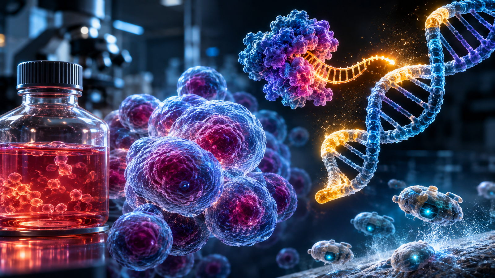
In Ridley Scott’s science fiction masterpiece Blade Runner (1982), the bioengineered replicant Roy Batty crosses a rain-drenched, dystopian Los Angeles to confront his creator, Eldon Tyrell. Nexus-6 replicants possess superhuman strength and agility, yet carry a deliberate curse written into their genetic code: an unyielding four-year lifespan. Conversely, in Marvel comics and cinema, the anti-hero Deadpool embodies the opposite extreme: his regenerative healing factor is driven by a hyperactive cancer whose cells never stop dividing, granting biological immortality at the cost of runaway cellular proliferation.
Beyond the silver screen, real human biology is governed by this exact tug-of-war between a programmed mortality clock and the danger of endless division. Nearly all of the trillions of cells in our body are born with a built-in countdown timer that limits how many times they can multiply before aging and dying.
Yet modern science has discovered that the boundary between cellular life and death is far more malleable than classical medicine ever imagined. On one side, the famous HeLa cells — harvested from a human patient in 1951 — bypassed the aging clock through the enzyme telomerase, remaining alive and continuously dividing for over seven decades in laboratories worldwide. On the other side, a breakthrough detailed in a landmark study published in the journal Physiology by biologists Peter Noble, Alex Pozhitkov, and Michael Levin unveiled the existence of a “Third State of Life”: when cells are removed from an organism (even after the individual’s death) and placed in a new environment, they can spontaneously reorganize into swimming biological robots — Xenobots and Anthrobots — capable of navigating microscopic spaces and healing damaged human neurons without any genetic modification.
In this accessible technical essay, we open the black box of cellular mortality and emancipation: you will visualize how the telomere clock works, the molecular trick that made HeLa cells eternal, and how living biobots are rewriting the concepts of life and death in regenerative medicine.
1. The Mortality Clock: Telomeres, Telomerase, and the Hayflick Limit
During the first half of the twentieth century, many scientists believed that any human cell cultured in a glass dish with proper nutrients would live forever. In 1961, American anatomist Leonard Hayflick overturned that myth by demonstrating that healthy human cells divide only 40 to 60 times before stopping permanently and entering a state of biological retirement called cellular senescence — a barrier now known as the Hayflick Limit.
The Shoelace Tip and the Bricklayer Problem
Why do our cells stop dividing after 40 to 60 rounds? The answer lies at the tips of our chromosomes and was deciphered by scientists Elizabeth Blackburn, Carol Greider, and Jack Szostak — an achievement that earned them the 2009 Nobel Prize in Physiology or Medicine, detailed in the historical review on telomeres and telomerase at PubMed Central.
To visualize this mechanism without complex biochemical formulas, picture two everyday images:
- The Plastic Tip of a Shoelace (Telomeres): Inside the nucleus of every cell, our DNA is coiled into 46 bundles called chromosomes. At the tip of each chromosome sits a protective cap made of thousands of repeats of the same six-letter chemical sequence (TTAGGG). These shields are telomeres: they work just like the hard plastic tip (aglet) at the end of a shoelace, preventing the DNA strand from fraying or sticking to other chromosomes.
- The Bricklayer Painting a Wall Backward (The End-Replication Problem): Every time a cell prepares to divide in two, a copying machine called DNA polymerase must duplicate the entire DNA strand. The catch is that this copier works like a bricklayer walking backward while painting the top of a wall: when he reaches the very last brick at the edge — right where his own feet are standing — he cannot paint the final spot beneath his boots.
In practical terms: with every cell division, the chromosome loses 50 to 200 “bricks” (base pairs) from its protective cap. At birth, a human cell has roughly 10,000 to 15,000 telomere bricks; after 50 divisions, the plastic tip wears away. Once actual genetic code is exposed, a safety sensor called the p53 protein pulls the emergency brake and forbids the cell from dividing further, protecting the body against mutated cells.
The Molecular Time Machine: How the Enzyme Telomerase Works
If every division shortens DNA, how do stem cells and embryos build an entire baby without being born old? They use a molecular “time machine” called Telomerase.
Telomerase acts like an extendable scaffold that attaches to the end of the wall and replaces the lost TTAGGG bricks after each division, resetting the countdown timer. In ordinary adult body cells (skin, liver, lungs), the gene that manufactures telomerase is intentionally switched off after birth: cellular aging is the evolutionary price we pay to prevent damaged cells from multiplying endlessly into tumors.
2. The Accidental Immortality of Henrietta Lacks: The Secret of HeLa Cells
In February 1951, a young African American woman named Henrietta Lacks was admitted to Johns Hopkins Hospital in Baltimore to treat an aggressive cervical tumor. Without her knowledge or consent — a standard practice at the time — a tissue sample was sent to researcher George Otto Gey, who had spent decades trying to keep human cells alive outside the body.
While all previous human cell cultures died within days, Henrietta’s cells — labeled HeLa from her initials — doubled every 24 hours without stopping. Henrietta passed away in October 1951 at age 31, but her cells became the first immortal human cell line in history.
Cut Brake Cables and a Jammed Accelerator: The HPV-18 Viral Hijack
More than sixty years later, the genomic and transcriptomic sequencing of the HeLa line revealed the molecular storm that rendered those cells eternal. Henrietta’s tumor had been triggered by Human Papillomavirus type 18 (HPV-18), which executed a dual sabotage worthy of a rogue mechanic:
- Cutting the Brake Cables (Viral Proteins E6 and E7): The virus produced two saboteur proteins named E6 and E7. Protein E6 hunted down and destroyed p53 (the guardian that halts damaged cells), while E7 disabled the retinoblastoma suppressor protein (pRb). Stripped of brakes, the cell lost its ability to halt division or undergo programmed suicide (apoptosis).
- Welding the Accelerator Pedal to the Floor (Telomerase Reactivation): The virus inserted its own genetic code into Henrietta’s chromosome 8, right next to a growth-promoting gene called c-MYC. That short-circuit switched the production of telomerase to maximum volume. With telomerase rebuilding the chromosomal caps after every division, the Hayflick Limit vanished.
The result of this unbraked division was extreme mutation: whereas a healthy human cell holds 46 organized chromosomes, a modern HeLa cell carries between 76 and 80 scrambled chromosomes, behaving almost like an autonomous laboratory microorganism.
The Impact of 50 Million Metric Tons on Global Medicine
Over seven decades, scientists have grown more than 50 million metric tons of HeLa cells in laboratories worldwide, underpinning over 110,000 scientific studies:
- The Polio Vaccine (1953): Jonas Salk used HeLa cell factories to mass-test the vaccine that saved millions of children from infantile paralysis.
- The Discovery of HPV Vaccines and the Nobel Prize: In the 1980s, virologist Harald zur Hausen discovered HPV-18 inside HeLa cells, proving that viruses cause cervical cancer and paving the way for modern HPV vaccines.
- In Vitro Fertilization, HIV Virology, and Space Biology: HeLa cells rode into space aboard early orbital missions before human astronauts to test how cosmic radiation and microgravity affect human tissues.
3. The Third State of Life: What Happens When the Body Dies, Yet the Cell Continues?
If HeLa cells proved that parts of a human can survive decades after the individual’s death, an even deeper question emerged in contemporary biology: what happens to healthy cells when the body dies, or when they are freed from their original organ?
In 2024, biologists Peter Noble and Alex Pozhitkov, alongside Michael Levin and researchers across major US universities, published the Physiology paper that christened this uncharted territory the “Third State of Life.”
Genes Awakening in the Twilight of Death
In traditional medicine, cardiac and brain death instantly terminates all organized bodily activity. Yet by analyzing 701 postmortem human tissue samples and monitoring animal models for up to 96 hours after cardiac arrest, researchers uncovered a startling phenomenon:
- Hundreds of Genes Wake Up After Death: Rather than all biological functions shutting down together, roughly 2% of genes surge in activity hours and days after clinical death, activating stress-response pathways, immunity, and — most intriguingly — embryonic development genes that had lain dormant since the individual was a fetus in the womb.
- Autonomous Survival of Fibroblasts: In human skin samples, roughly 50% of structural cells (fibroblasts) continued communicating via chemical signals as if unaware that the host body had died.
The explanation proposed by the authors redefines biology: during life, trillions of cells obey a central “bioelectric network” that compels them to work as liver, skin, or lung. When the organism dies (or when cells are isolated in a dish), that central management disappears. Freed from bodily hierarchy, surviving cells attempt to reboot their collective life from scratch — entering the Third State.
4. Xenobots and Anthrobots: Engineering Living Robots Without Genetic Editing
Definitive proof of this Third State came to life in the laboratories of Michael Levin (Tufts University and Harvard’s Wyss Institute) and Josh Bongard (University of Vermont), connecting directly to the programmable biology ecosystem we explore in The Definitive Guide to Biotechnology: From CRISPR and mRNA Vaccines to Neuralink and Longevity.
From Amphibian Xenobots to 100% Human Anthrobots
In 2020, as documented in the original Xenobots research in the journal PNAS, scientists harvested embryonic skin cells from the African clawed frog Xenopus laevis and allowed them to reassemble in vitro. Instead of forming a tadpole or a flat sheet of skin, the cells organized into tiny living spheres that swam across the dish and even pushed loose cells together to assemble new copies of themselves (kinematic self-replication).
A crucial question remained: could cells from adult human beings — with no embryonic tricks and zero genetic editing — do the same?
The answer arrived with the creation of Anthrobots, led by researcher Gizem Gumuskaya and Michael Levin and published in the seminal Anthrobots study in Advanced Science.
The Inside-Out Rowboat Analogy
To understand how scientists turned a human lung cell into a swimming robot without altering a single letter of DNA, picture the following visual analogy:
Inside our windpipe (trachea) and bronchi, epithelial cells stand side by side like workers bolted to the floor, using thousands of microscopic hairs (called cilia) to sweep dust and mucus out of the lungs. When grown inside a thick gel in the laboratory, they form a hollow ball with all those hairs pointing inward — like a boat where all the rowers are rowing inside a closed hull, going nowhere.
Gumuskaya and Levin’s engineering trick was purely environmental: after 14 days in thick gel, they dissolved the matrix and transferred the spheres into a low-viscosity liquid. Sensing the fluid change around them, the cells flipped their architecture inside out within 7 days: they pushed all their cilia to the outside of the hull, just like the oars of a Roman galley, and began swimming through the liquid at speeds of 5 to 50 micrometers per second.
Comparative Table: The Three Regimes of Cellular Life
The matrix below compares an ordinary adult human cell, an immortal HeLa cell, and a Third-State human biobot side by side:
| Biological Dimension | Ordinary Adult Human Cell | Immortal HeLa Cell (Cancer / HPV-18) | Human Biobot (Anthrobot — 3rd State) |
|---|---|---|---|
| Tissue Origin | Differentiated organs of the living body | Cervical tumor of Henrietta Lacks (1951) | Adult human tracheal cells with zero gene editing |
| Genetic Code & Chromosomes | 46 intact human chromosomes | 76 to 80 scrambled chromosomes with viral DNA | 46 original chromosomes from the patient |
| Division Clock (Telomerase) | Switched off; halts after 40–60 divisions (Hayflick Limit) | Hyperactive telomerase and no p53 brake; endless division | Safe, self-limiting lifespan of 45 to 60 days |
| Behavior & Mobility | Static, locked into the organ’s native architecture | Uncontrolled, invasive proliferation | Swims autonomously using external cilia in loops or lines |
| Medical Application | Normal physiological maintenance | Mass testing of vaccines, drugs, and viruses | Stitching injured neurons and delivering targeted therapy |
5. Epigenetic Rejuvenation and the Healing of Human Neurons
The most astonishing behavior of Anthrobots emerged when scientists placed these tiny swimmers (measuring between the width of a human hair and the tip of a sharpened pencil) onto a laboratory sheet of human neurons that had been torn open by a deep mechanical scratch.
“Superbot Bridges”: Stitching Neural Wounds in 72 Hours
Even though they are made solely of tracheal airway cells — which never interact with brain tissue in nature — when clusters of Anthrobots encountered the gap in the neurons, they gathered over the wound to form a living “superbot bridge.” Within just three days (72 hours) beneath that bridge of biobots, the severed human neurons regrew and completely closed the neural scar, requiring no external drugs.
Resetting the Aging Clock Across 9,000 Genes
Why do adult airway cells acquire locomotion and healing powers upon entering the Third State? In 2025, a follow-up study by the Tufts team published in the investigation on the life cycle and rejuvenation of Anthrobots sequenced the internal RNA of the biobots and uncovered a profound transformation:
- Spontaneous Reprogramming of Half the Genome: Without any genetic modification, Anthrobot cells spontaneously altered the expression of over 9,000 genes (nearly half of the human genome), reactivating ancient developmental programs for symmetry and tissue repair.
- Reversal of Biological Age: By measuring the epigenetic methylation clock of the cells before and after becoming Anthrobots, scientists discovered that assembling into a biobot turns back the cells’ biological age compared to their adult source tissue.
- Programmed Safe Biodegradation: Unlike HeLa cells — which are cancerous and multiply forever — Anthrobots do not reproduce uncontrollably: after fulfilling their mission over 45 to 60 days, they naturally biodegrade, acting as temporary, disposable surgical robots.
6. Critical Analysis, Bioethics, and the Future of Regenerative Medicine
From Henrietta Lacks’s legacy to Michael Levin’s biobots, manipulating the boundary between cellular life and death carries extraordinary clinical promise, yet also confronts technical bottlenecks and ethical dilemmas that science must address transparently.
Four Technical Bottlenecks and Bioethical Dilemmas at the Cellular Frontier
Bringing these technologies into hospitals depends on overcoming four real-world barriers:
- The Telomerase Paradox (Longevity vs. Cancer): Why don’t we inject telomerase across the human body to live forever like HeLa cells? Because indiscriminately reactivating telomerase in adult tissues that have accumulated lifelong DNA mutations risks fueling silent tumors. Modern longevity therapies aim to activate telomerase or epigenetic factors only in short, controlled pulses without disabling the p53 guardian.
- Cross-Contamination by HeLa Cells in Laboratories: The proliferative aggressiveness of HeLa cells is so extreme that a single cell carried on an aerosol droplet or unwashed glove can invade neighboring culture dishes and ruin months of research. Between 10% and 20% of cell lines in global repositories have been accidentally overgrown and replaced by HeLa cells.
- Navigating Anthrobots Inside the Living Human Body: While Anthrobots swim and heal neurons across flat laboratory dishes, steering them through the three-dimensional labyrinth of blood vessels, arterial flow, and mucus barriers inside a living patient will require external bioelectric or magnetic guidance systems.
- Genomic Consent and the Moral Status of Biobots: The story of Henrietta Lacks — whose cells generated billions of dollars for the biomedical industry while her family remained uncompensated for decades, leading to landmark NIH agreements in 2013 and biotech legal settlements in the 2020s — reshaped global tissue-consent laws. Now, Third-State biobots raise a new regulatory question: how should law classify living human constructs that are neither embryos nor organs, but autonomous biological machines?
Life Beyond Evolution’s Default Manual
The journey that began in 1951 with the accidental immortality of HeLa cells and reached Third-State Anthrobots in 2026 reveals a profound truth: human DNA is not a rigid, single-purpose script, but a versatile toolkit capable of building new forms of life when given the right environmental cues.
In the near future, rather than swallowing synthetic pills that trigger side effects across the entire body, a physician may take a simple swab of cells from your own throat, transform them into thousands of personalized Anthrobots — completely invisible to your immune system — and deploy them temporarily to clear arterial plaque, hunt down hidden cancer cells, or bridge severed nerves in a damaged spinal cord.
Facing this frontier where our own cells can be turned into tiny living healer robots, which medical application do you consider most urgent for the coming decades: regenerating spinal and brain injuries with biobots, or safely mastering telomerase to slow human aging? Share your perspective in the comments and pass this article along!
🔭 Continue Exploring the Frontier of Science and Technology: